
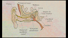
The Ear

The ears perform two sensory functions, hearing and maintenance of body balance. Anatomically, the ear can be divided into three major sections called the outer ear, the middle ear and the inner ear . The outer ear consists of the pinna and external auditory meatus (canal). The pinna collects the vibrations in the air which produce sound. external nal auditory The meatus leads inwards and extends up to the tympanic membrane (the ear drum). There are very fine hairs and wax-secreting glands in the skin of the pinna and the meatus. The tympanic membrane is composed of connective tissues covered with skin outside and with mucus membrane inside.
The middle ear contains three ossicles called malleus, incus and stapes which are attached to one another in a chain-like fashion. The malleus is attached to the tympanic membrane and the stapes is attached to the oval window of the cochlea. The ear ossicles increase the efficiency of transmission of sound waves to the inner ear. An Eustachian tube connects the middle ear cavity with the pharynx. The Eustachian tube helps in equalising the pressures on either sides of the ear drum.
The fluid-filled inner ear called labyrinth consists of two parts, the bony and the membranous labyrinths. The bony labyrinth is a series of channels. Inside these channels lies the membranous labyrinth, which is surrounded by a fluid called perilymph. The membranous labyrinth is filled with a fluid called endolymph. The coiled portion of the labyrinth is called cochlea. The membranes constituting cochlea, the reissner's and basilar, divide the surounding perilymph filled bony labyrinth into an upper scala vestibuli and a lower scala tympani.The space within cochlea called scala media is filled with endolymph. At the base of the cochlea, the scala vestibuli ends at the oval window, while the scala tympani terminates at the round window which opens to the middle ear.
The organ of corti is a structure located on the basilar membrane which contains hair cells that act as auditory receptors. The hair cells are present in rows on the internal side of the organ of corti. The basal end of the hair cell is in close contact with the afferent nerve fibres. A large number of processes called stereo cilia are projected from the apical part of each hair cell. Above the rows of the hair cells is a thin elastic membrane called tectorial membrane.
The inner ear also contains a complex system called vestibular apparatus, located above the cochlea. The vestibular apparatus is composed of three semi-circular canals and the otolith (macula is the sensory part of saccule and utricle). Each semi-circular canal lies in a different plane at right angles to each other. The membranous canals are suspended in the perilymph of the bony canals.
The base of canals is swollen and is called ampulla, which contains a projecting ridge called crista ampullaris which has hair cells. The saccule and utricle contain a projecting ridge called macula. The crista and macula are the specific receptors of the vestibular apparatus responsible for maintenance of balance of the body and posture.
Mechanism of Hearing
How does ear convert sound waves into neural impulses, which are sensed and processed by the brain enabling us to recognise a sound? The external ear receives sound waves and directs them to the ear drum. The ear drum vibrates in response to the sound waves and these vibrations are transmitted through the ear ossicles (malleus, incus and stapes) to the oval window. The vibrations are passed through the oval window on to the fluid of the cochlea, where they generate waves in the lymphs.
The waves in the lymphs induce a ripple in the basilar membrane. These movements of the basilar membrane bend the hair cells, pressing them against the tectorial membrane. As a result, nerve impulses are generated in the associated afferent neurons. These impulses are transmitted by the afferent fibres via auditory nerves to the auditory cortex of the brain. where the impulses are analysed and the sound is recognised.
Thanks for Reading My Article ☺️ . If You Have Any Problem Then Contact Me.


