
MUSCLES

The cilia and flagella are the outgrowths of the cell membrane. Flagellar movement helps in the swimming a spermatozoa. hance of water current in the canal system of sponge maintenance and in locomotion of Protozoans like Euglena. Muscle is a specialisee tissue of mesodermal origin. About 40-50 per cent of the bod- weight of a human adult is contributed by muscles. They hav special properties like excitability, contractility, extensibility ane elasticity. Muscles have been classified using different criteria namely location, appearance and nature of regulation of thei activities. Based on their location, three types of muscles ar identified: (i) Skeletal (ii) Visceral and (iii) Cardiac.
Skeletal muscles are closely associated with the skeletal component of the body. They have a striped appearance under the microscope ane hence are called striated muscles. As their activities are under the voluntary control of the nervous system, they are known as voluntar muscles too. They are primarily involved in locomotory actions and changes of body postures.
Visceral muscles are located in the inner walls of hollow visceral organ of the body like the alimentary canal, reproductive tract, etc. They do no exhibit any striation and are smooth in appearance. Hence, they are calle smooth muscles (nonstriated muscle). Their activities are not under th voluntary control of the nervous system and are therefore known a involuntary muscles. They assist, for example, in the transportation of food through the digestive tract and gametes through the genital tract.
As the name suggests, Cardiac muscles are the muscles of heart Many cardiac muscle cells assemble in a branching pattern to form cardiac muscle. Based on appearance, cardiac muscles are striated. The are involuntary in nature as the nervous system does not control the activities directly.
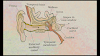
Let us examine a skeletal muscle in detail to understand the structur and mechanism of contraction. Each organised skeletal muscle in ou body is made of a number of muscle bundles or fascicles held together by a common collagenous connective tissue layer called fascia. Eas muscle bundle contains a number of muscle fibres. Each muscle fibre is lined by the plasma membrane called sarcolemma enclosing the sarcoplasm. Muscle fibre is a syncitium as the sarcoplasm contains many nuclei. The endoplasmic reticulum, i.e., sarcoplasmic reticulum of the muscle fibres is the store house of calcium ions. A characteristic feature of the muscle fibre is the presence of a large number of parallelly arranged filaments in the sarcoplasm called myofilaments or myofibrils. Each myofibril has alternate dark and light bands on it. A detailed study of the myofibril has established that the striated appearance is due to the distribution pattern of two important proteins - Actin and Myosin. The light bands contain actin and is called I-band or Isotropic band, whereas the dark band called 'A' or Anisotropic band contains myosin. Both the proteins are arranged as rod-like structures, parallel to each other and also to the longitudinal axis of the myofibrils. Actin filaments are thinner as compared to the myosin filaments, hence are commonly called thin and thick filaments respectively. In the centre of each 'T' band is an elastic fibre called 'Z' line which bisects it. The thin filaments are firmly attached to the 'Z' line. The thick filaments in the A' band are also held together in the middle of this band by a thin fibrous membrane called 'M' line. The 'A' and T' bands are arranged alternately throughout the length of the myofibrils. The portion of the myofibril between two successive 'Z' lines is considered as the functional unit of contraction and is called a sarcomere (Figure 20.2). In a resting state, the edges of thin filaments on either side of the thick filaments partially overlap the free ends of the thick filaments leaving the central part of the thick filaments. This central part of thick filament, not overlapped by thin filaments is called the 'H' zone.
Thanks for Reading My Article ☺️ . If You Have Any Problem Then Contact Me.


