

Skeletal system consists of a framework of bones and a few cartilages. This system has a significant role in movement shown by the body. Imagine chewing food without jaws bones and walking around without the limb bones. Bone and cartilage are specialised connective tissues. The former has a very hard Matrix due to calcium salts in it and the latter has slightly pliable Matrix due to chondroitin salts. In human beings, this system is made up of 206 bones and a few cartilages. It is grouped into two principal divisions - the axial and the appendicular skeleton.
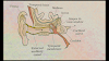
Axial skeleton comprises 80 bones distributed along the main axis of the body. The skull, vertebral column, sternum and ribs constitute axial skeleton. The skull is composed of two sets of bones cranial and facial, that total to 22 bones. Cranial bones are 8 in number. They form the hard protective outer covering, cranium for the brain. The facial region is made up of 14 skeletal elements which form the front part of the skull. A single U - shaped bone called hyoid is present at the base of the buccal cavity and it is also included in the skull. Each middle ear contains three tiny bones - Malleus, Incus and stapes, collectively called Earlier Ossicles. The skull region articulates with the superior region of the vertebral column with the help of two occipital condyles (dicondylic skull ).

Our vertebral column is formed by 26 serially arranged units called vertebrae and is dorsally placed. It extends from the base of the skull and constitutes the main framework of the trunk. Each vertebral has a central hollow portion (neural canal ) through which the spinal cord phases. First vertebra is the atlas and it articulates with the occipital condyles. The vertebral column is differentiated into cervical (7), thoracic (12), lumber (5), sacral (1-fused) and coccygeal (1-fused) regions starting from the skull. The number of cervical vertebrae are seven in almost all the mammals including humain beings. The vertebral column protects the spinal cord, supports the head and serves as the point of attachment for the ribs and musculature of the back. Sternum is a flat bone on the ventral midline of thorax.

There are 12 pairs of ribs. Each rib is a thin flat bone connected dorsally to the vertebral column and ventrally to the sternum. It has two articulation surfaces on its dorsal end and is hence called bicephalic. First seven pairs of ribs are called true ribs. Dorsally, they are attached to the thoracic vertebrae and ventrally connected to the sternum with the help of hyaline cartilage. The 8th, 9th and 10th pairs of ribs do not articulate directly with the sternum but join the seventh rib with the help of hyaline cartilage. These are called vertebrochondral (false) ribs. Last 2 pairs (11th and 12th) of ribs are not connected ventrally and are therefore, called floating ribs. Thoracic mn vertebrae, ribs and sternum together form the rib cage.
The bones of the limbs alongwith their girdles constitute the appendicular skeleton. Each limb is made of 30 bones. The bones of the hand (fore limb) are humerus, radius and ulna, carpals (wrist bones 8 in number), metacarpals (palm bones - 5 in number) and phalanges (digits 14 in number) (Figure 20.9). Femur (thigh bone - the longest bone). tibia and fibula, tarsals (ankle bones - 7 in number), metatarsals (5 in number) and phalanges (digits 14 in number) are the bones of the legs (hind limb). A cup shaped bone called patella cover the knee ventrally (knee cap).


Thanks for Reading My Article ☺️ . If You Have Any Problem Then Contact Me.


